

Hypercholesterolemia or high cholesterol is often asymptomatic until it has reached an advanced stage, but the typical ocular manifestations include xanthelasma and arcus corneae, which are considered early signs of dyslipidemia and correlate with increased cardiovascular risk. Their development is as a result of lipid deposits into ocular tissues; thus, ocular findings may warrant investigation into the lipid profile of apparently asymptomatic individuals. This early detection allows for the institution of appropriate measures to help reduce the risk of further atherosclerosis and maintain good vision.
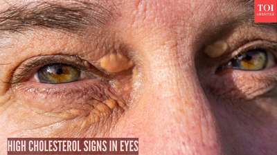
Xanthelasma: Cholesterol deposits periorbitally
Xanthelasma appears as soft, yellowish plaques on the medial eyelids or nasojugal fold, the result of cholesterol extravasation into dermal macrophages. These lesions develop insidiously, with no associated visual disturbance or discomfort unless they are enlarged, but they are strongly associated with hyperlipidemia, hypothyroidism, or diabetes mellitus. The prevalence increases in women over 40 years, particularly postmenopausal, because the decline of estrogen levels exacerbates lipid dysregulation. Management includes treatment options such as trichloroacetic acid ablation, laser therapy, and excision for cosmetic correction, together with systemic lipid management.
Corneal Arcus: Annular lipid infiltration

Arcus corneae appears as a circular, white-to-grayish opacity at the corneoscleral limbus-and is made up of lipid-laden keratocytes. When it is called arcus senilis in individuals above 60 years, it is benign. However, when it presents as arcus lipoides in individuals below 40 years, this indicates familial hypercholesterolemia or early vascular disease. The pathology carries no refractive error or symptomatic consequence but requires extensive lipid screening and stratification for cardiovascular risk.
Hollenhorst Plaques : Retinal emboli
Hollenhorst plaques are refractile, yellow cholesterol crystals within retinal arterioles that arise from proximal atherosclerotic plaques, often from the carotid. Visualized solely through dilated pupillary funduscopy, these emboli induce transient branch retinal artery occlusion, which can become permanent ischemia if not resolved. They also portend systemic embolization potential and mandate urgent neuroimaging and antiplatelet therapy.
Retinal vascular occlusions
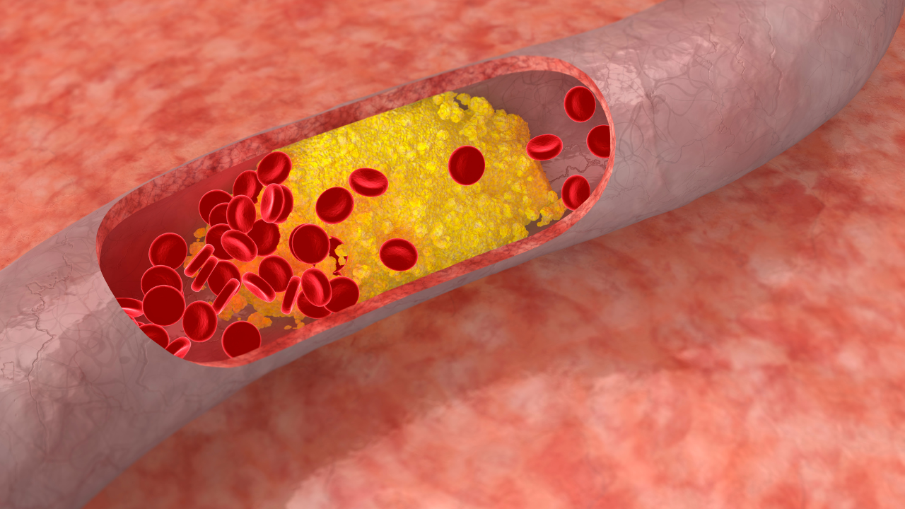
Ischemic Sequelae Central or branch RVO or RAO results from thrombosis caused by hypercholesterolemic atheroma and causes an acute monocular loss of vision, relative afferent pupillary defect-and retinal hemorrhages on ophthalmoscopy. RVO is more common and presents with cotton-wool spots and venous dilation, whereas RAO presents with pale retina and cherry-red foveal reflex. These are ocular emergencies and require the administration of intravitreal anti-VEGF, thrombolysis, or panretinal photocoagulation to prevent neovascular complications.
Pathophysiological rationale for ocular involvement

Thin basement membranes and high metabolic demand in ocular vasculature predispose it to LDL oxidation and foam cell formation, well before systemic manifestations. Dietary saturated fats, sedentary behavior, diabetes, and obesity are risk amplifiers, prevalent in South Asian cohorts similar to vitamin D-deficient populations. Postmenopausal hormonal changes further elevate LDL in women.
Diagnostic recommendations
Diagnosis is established with annual comprehensive ophthalmoscopy with lipid panel-total cholesterol >200 mg/dL, LDL >130 mg/dL indicative. Lifestyle modifications include a Mediterranean diet rich in omega-3s, aerobic exercise (150 minutes a week)-and smoking cessation. Statins achieve target LDL <100 mg/dL in high-risk cases. Elective xanthelasma remediation; vascular occlusions require multidisciplinary intervention.